T1 Vertebrae - Each thoracic spinal nerve is named for the vertebra above it.. However, correlation with the lesion appearances on other mr imaging sequences and imaging modalities as well as with the clinical history may occasionally suggest otherwise. The t1 vertebra is the first (uppermost) of the twelve (12) thoracic vertebrae that make up the central and largest section of the spinal column between the lumbar vertebrae below and the cervical vertebrae above. Simply line up the vertebral level with the possible symptoms and you will see some surprising connections of symptoms that relate to your spine. Each thoracic spinal nerve is named for the vertebra above it. The thoracic spinal vertebrae consist of 12 total vertebrae and are located between the cervical vertebrae (which begin at the base of the skull) and the lumbar spinal vertebrae.
It is located between the cervical and lumbar levels, right in the middle of the spine at your mid back. T1 also has a spinous process more horizontal than other thoracic vertebrae. They are very common and occur in approximately 10 percent of the world's population. The t1 nerve roots affect sensation in your inner forearm and the ability to spread your fingers apart. Hemangiomas most often appear in adults between the ages of 30 and 50.
Thoracic Vertebrae Anatomy Function And Definition Kenhub from i.vimeocdn.com Simply line up the vertebral level with the possible symptoms and you will see some surprising connections of symptoms that relate to your spine. If your fingers are over the c7 and t1 sps, the c7 sp will move while the t1 sp stays relatively fixed. The t1 vertebra is the first of twelve vertebrae of the thoracic spinal column. Eg the t4 nerve root runs between the t4 vertebraand t5 vertebra. However, correlation with the lesion appearances on other mr imaging sequences and imaging modalities as well as with the clinical history may occasionally suggest otherwise. The t2 vertebra is the second vertebra in the thoracic spinal column. Since each descending vertebra is larger than the one before it to support the greater weight, thoracic vertebra t1 is the smallest vertebra of the thoracic region. T1 is an atypical thoracic vertebra.
The vast majority of t1 hyperintense vertebral column lesions are benign.
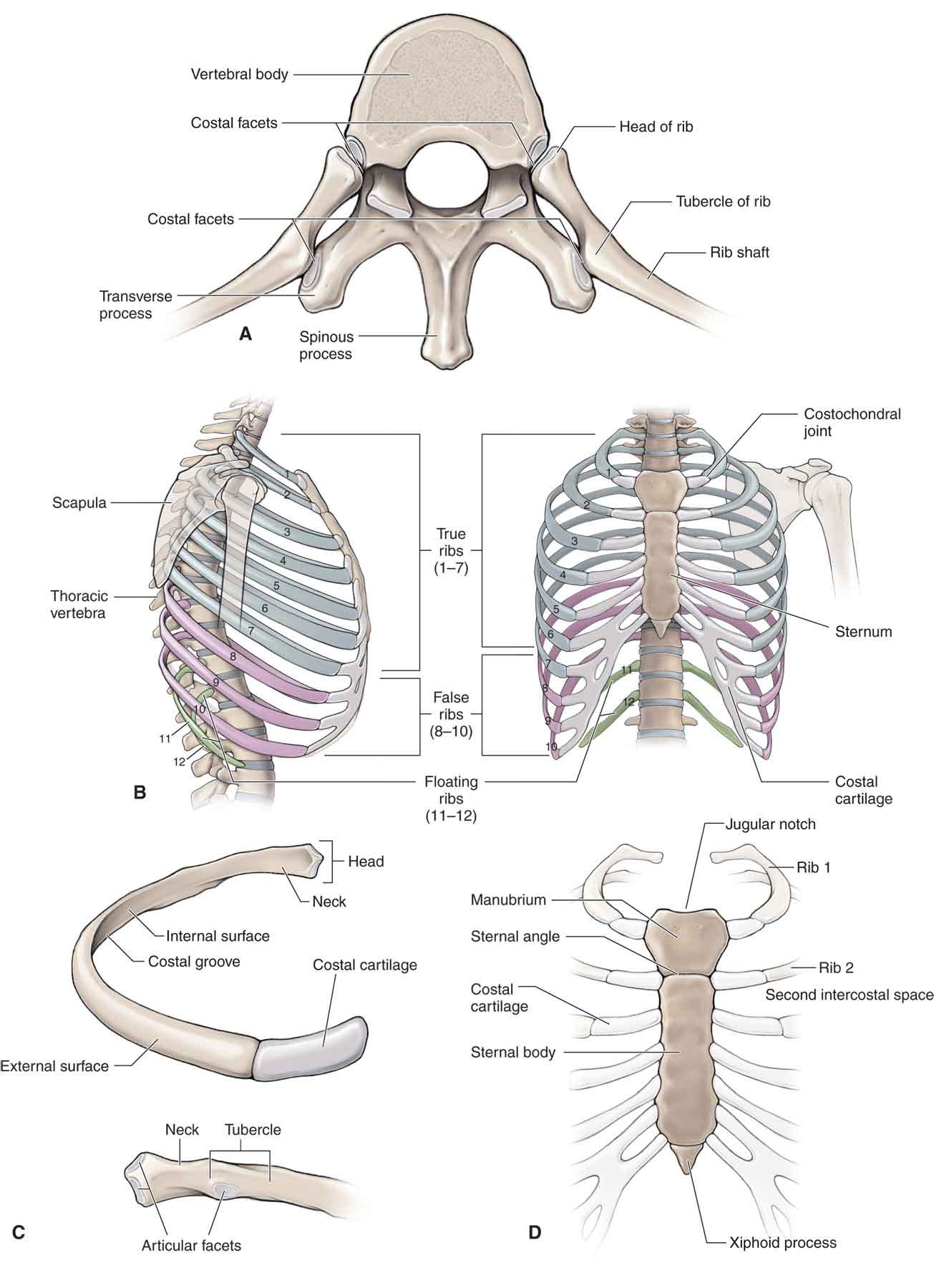
If your fingers are over the c7 and t1 sps, the c7 sp will move while the t1 sp stays relatively fixed. It is quite possible for survivors with spinal cord injuries at this level to live independently. The thoracic spine has 12 nerve roots (t1 to t12) on each side of the spine that branch from the spinal cord and control motor and sensory signals mostly for the upper back, chest, and abdomen. A vertebral tumor is a type of spinal tumor affecting the bones or vertebrae of the spine. It contains lips on the upper surface of the body. The thoracic spine is made up of twelve vertebrae, known as t1 through t12. The thorax (chest) is a cage of. While larger than the c7 vertebra above it, the t1 is the smallest of the thoracic vertebrae. They are characterized by small pedicles, long spinous processes, and relatively large intervertebral foramen (neural passageways), which result in less incidence of nerve compression. T3, t4, and t5 nerve roots affect sensation at the upper chest and back. Tumors that affect the vertebrae have often spread (metastasized) from cancers in other parts of the body. On the chart below you will see 4 columns (vertebral level, nerve root, innervation, and possible symptoms). T2 nerve roots affect sensation around the armpits and upper chest.
T1 is an atypical thoracic vertebra. Tumors that affect the vertebrae have often spread (metastasized) from cancers in other parts of the body. Bones and cartilage (a type of tissue) that extends from the. If your fingers are over the c7 and t1 sps, the c7 sp will move while the t1 sp stays relatively fixed. Hemangiomas most often appear in adults between the ages of 30 and 50.
The Thoracic Spine Musculoskeletal Key from musculoskeletalkey.com It is quite possible for survivors with spinal cord injuries at this level to live independently. T2 the second vertebra in the thoracic spine is responsible for helping to support the rib cage. To test for individual vertebra start by finding t1. T1 (1st thoracic vertebra) the t1 vertebra is the first (uppermost) of the twelve (12) thoracic vertebrae that make up the central and largest section of the spinal column between the lumbar vertebrae below and the cervical vertebrae above. They are very common and occur in approximately 10 percent of the world's population. Simply line up the vertebral level with the possible symptoms and you will see some surprising connections of symptoms that relate to your spine. Form an opening in which the spinal cord passes) that are. Each thoracic spinal nerve is named for the vertebra above it.
T1 is an atypical thoracic vertebra.
To test for individual vertebra start by finding t1. Hemangiomas most often appear in adults between the ages of 30 and 50. Spinal tumors that begin within the spinal cord or the covering of the spinal cord (dura) are called spinal cord tumors. The vertebrae themselves are numbered in descending order, and t1 is located at the top of the thoracic spine. The vast majority of t1 hyperintense vertebral column lesions are benign. Eg the t4 nerve root runs between the t4 vertebraand t5 vertebra. T3, t4, and t5 nerve roots affect sensation at the upper chest and back. In contrast to typical thoracic vertebrae, it contains a complete facet for the 1 st rib and a demifacet for the 2 nd rib. Bottom of the neck to a muscle known as the. These vertebrae are connected by a pair of facet joints in the back and each has a vertebral body, 2 pedicles, 2 transverse processes (bony humps on the side where muscles can attach and pull on the vertebrae), 2 lamina, and a spinous process. The cervical spine (neck), the thoracic spine (where your ribs attach), the lumbar spine, and finally the sacrum and coccyx. Form an opening in which the spinal cord passes) that are. It is located between the cervical and lumbar levels, right in the middle of the spine at your mid back.
Behind an area of the body known as the thorax, which is. The t1 vertebra is the first of twelve vertebrae of the thoracic spinal column. For each of the 12 thoracic vertebrae, there is a corresponding pair of ribs attached to them. T1 is an atypical thoracic vertebra. It contains lips on the upper surface of the body.
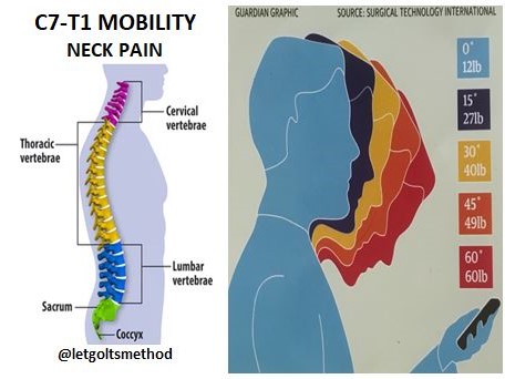
Thoracic Spine from letgoltsmethod.com What is the t2 vertebra? To test for individual vertebra start by finding t1. Tumors that affect the vertebrae have often spread (metastasized) from cancers in other parts of the body. Eg the t4 nerve root runs between the t4 vertebraand t5 vertebra. Bottom of the neck to a muscle known as the. They are characterized by small pedicles, long spinous processes, and relatively large intervertebral foramen (neural passageways), which result in less incidence of nerve compression. They are very common and occur in approximately 10 percent of the world's population. Bones and cartilage (a type of tissue) that extends from the.
Each thoracic spinal nerve is named for the vertebra above it.
T1 is an atypical thoracic vertebra. Spinal tumors that begin within the spinal cord or the covering of the spinal cord (dura) are called spinal cord tumors. Form an opening in which the spinal cord passes) that are. It is located between the cervical and lumbar levels, right in the middle of the spine at your mid back. While larger than the c7 vertebra above it, the t1 is the smallest of the thoracic vertebrae. Since each descending vertebra is larger than the one before it to support the greater weight, thoracic vertebra t1 is the smallest vertebra of the thoracic region. A vertebral tumor is a type of spinal tumor affecting the bones or vertebrae of the spine. Simply line up the vertebral level with the possible symptoms and you will see some surprising connections of symptoms that relate to your spine. The vast majority of t1 hyperintense vertebral column lesions are benign. They are very common and occur in approximately 10 percent of the world's population. However, correlation with the lesion appearances on other mr imaging sequences and imaging modalities as well as with the clinical history may occasionally suggest otherwise. Survivors with an injury at this level can expect to have the use of the head, neck, shoulders, arms, hands, and fingers. These facets are located across both the vertebral body and the pedicle.
While larger than the c7 vertebra above it, the t1 is the smallest of the thoracic vertebrae t1. These twelve vertebrae and nerves connect directly to the rib cage, thus allowing easy communication with the central parts of the body.


0 Komentar